Spinal Fusion – Fact Sheet

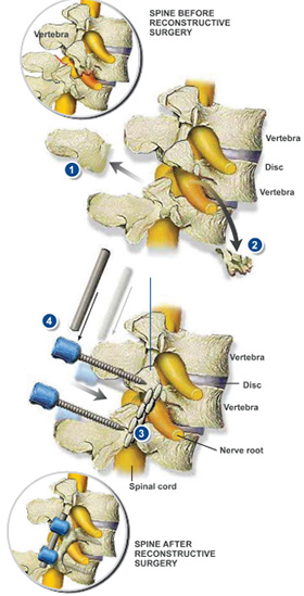
During this procedure, the surgeon permanently joins two or more vertebrae in the spine. The vertebrae will grow together to form a single, solid bone. Spinal fusion is commonly performed in the neck and lower back, and may be used to correct a wide range of problems in the spine. This animation shows a fusion in the lumbar spine to correct a condition called spondylolisthesis, in which weakened joints or fractured bones have allowed a vertebra to slip forward and pinch a nerve root. Preparation In preparation for the procedure, the patient is positioned and general anesthesia is administered. The surgeon creates an incision to access the lumbar spine.
Laminectomy
The surgeon removes the lamina, the protrusion at the rear of the vertebra. This is the portion of the vertebra that covers the nerve roots. Removing the lamina creates more space for the nerve roots.
Bone Cleared
Next, the surgeon clears any bone or debris that may be pressing against the nerve roots. This relieves pressure and pain.
Bone Grafts Implanted
The surgeon places bone grafts against the vertebrae. The bone grafts may be harvested from the patient’s own body, typically from the pelvis. They may also be taken from another donor.
Screws/Rods Inserted
The surgeon inserts hardware to hold the vertebrae together. The surgeon may utilize screws and rods or plates.
End of Procedure
When the procedure is complete, the incision is closed and bandaged. The patient may be placed in a back brace to restrict movement of the spine. The patient will be able to leave the hospital after two or three days. As the spine heals, the bone grafts will fuse with the vertebrae to create a solid, stable mass of bone

 Menu
Menu